Abstract
Momordica cochinchinensis (Dumbaya) is a local plant with high nutritional and medicinal potential, but its propagation is constrained by hard seed coats and low natural regeneration capacity. Tissue culture via callus induction offers an alternative strategy to support efficient propagation and further in vitro studies. This study aimed to develop a hypocotyl-derived callus induction protocol for M. cochinchinensis using different concentrations of 2,4-dichlorophenoxyacetic acid (2,4-D) and 6-benzylaminopurine (6-BA). The scientific contribution of this study lies in establishing a preliminary callus induction protocol using hypocotyl explants of Dumbaya, a local germplasm that has received limited attention in in vitro culture studies. Hypocotyl segments from four-week-old in vitro seedlings were cultured for eight weeks on Murashige and Skoog medium supplemented with three plant growth regulator combinations: A1, 2.0 mg L-1 2,4-D + 0.5 mg L-1 6-BA; A2, 1.5 mg L-1 2,4-D + 0.75 mg L-1 6-BA; and A3, 1.0 mg L-1 2,4-D + 1.5 mg L-1 6-BA. All tested treatments induced friable callus. Treatment A3 showed a trend toward higher fresh weight and more favorable yellowish white callus morphology, although the differences among treatments were not statistically significant. These findings indicate that 1.0 mg L-1 2,4-D combined with 1.5 mg L-1 6-BA may serve as a preliminary starting formulation for producing hypocotyl-derived callus of M.cochinchinensis for further regeneration, conservation, and secondary metabolite-related studies.
-
Key words: Momordica cochinchinensis, Hypocotyl explant, Plant tissue culture, Auxin-cytokinin interaction, Dumbaya germplasm
Introduction
Medicinal and nutraceutical plants are increasingly valued as sources of bioactive compounds for food, health, and pharmaceutical applications. Plant tissue culture has become an important enabling technology for supporting the propagation and biotechnological utilisation of these plants because in vitro systems can provide disease-free propagules, uniform planting material, and controlled platforms for the production of valuable metabolites (
Hussain et al. 2012). Among tissue culture approaches, callus culture is particularly useful for studying morphogenesis, genetic variability, and secondary-metabolite biosynthesis, while also serving as starting material for plant regeneration and genetic transformation (
Bojko et al. 2024;
Kruglova et al. 2023).
Momordica cochinchinensis (Dumbaya), a member of the Cucurbitaceae, has high nutritional and medicinal potential, particularly because of its carotenoid rich aril and reported antioxidant and pharmacological properties. However, the wider utilisation, domestication, and research development of this species are constrained by hard seed coats, erratic germination, and low natural regeneration capacity. These limitations reduce the availability of uniform plant material for cultivation, conservation, and experimental studies. Similar constraints related to seed-based propagation and environmental effects on seed quality have been reported in other medicinal crops, such as
Bupleurum chinense (
Li et al. 2023). Therefore, tissue-culture-based propagation offers a promising strategy to support the sustainable utilisation of
M. cochinchinensis by enabling rapid multiplication of selected genotypes and providing a foundation for downstream organogenesis and metabolite-related studies.
The interaction between explant type, genotype, culture medium, and plant growth regulators strongly influences callus induction. In particular, the balance between auxins and cytokinins is critical in determining callus formation, proliferation, morphology, and morphogenetic competence (
Bidabadi et al. 2020;
Kruglova et al. 2023). Auxins such as 2,4-D are widely used to promote cell dedifferentiation and callus initiation, whereas cytokinins such as BA support cell division and organogenic responses (
Andaryani et al. 2019;
Muthi’ah et al. 2023). Previous studies in diverse plant species have shown that 2,4-D and 6-BA combinations can markedly affect callus induction frequency, biomass accumulation, color, and texture, including in
Jatropha curcas (
Andaryani et al. 2019),
Calotropis gigantea (
Muthi’ah et al. 2023), and
Heritiera javanica (
Iswanto et al. 2025). However, inappropriate hormone combinations may cause poor callus proliferation, browning, compact callus formation, or reduced regeneration potential, as reported in callus cultures of
B. chinense and
Tussilago farfara (
Bojko et al. 2024;
Li et al. 2023).
In addition to hormonal balance, explant specificity is a key determinant of successful callus induction. Hypocotyl explants have been reported as responsive tissues for callus induction and regeneration in several medicinal and horticultural species. For example, hypocotyl-derived callus of
Nigella damascena showed high induction frequency and maintained regeneration capacity over extended culture periods (
Klimek-Chodacka et al. 2020), while hypocotyl callus of
B.chinense displayed superior shoot regeneration compared with other explants (
Li et al. 2023). These findings suggest that hypocotyls may also represent a suitable explant source for
M. cochinchinensis, particularly when combined with an appropriate auxin cytokinin regime. Nevertheless, the response of Dumbaya hypocotyls to defined 2,4-D and 6-BA concentrations has not yet been systematically evaluated.
Callus color and texture are important practical indicators of callus quality, vigour, and potential developmental competence. High-quality, friable, and light colored callus is generally associated with active cell proliferation and favourable regeneration potential, whereas dark, compact, or browning callus may indicate stress responses, phenolic accumulation, or reduced morphogenetic capacity (
Bojko et al. 2024;
Febriani et al. 2025). Studies on ornamental, medicinal, and horticultural species, including
Syzygium cumini, Aglaonema ‘Siam Aurora’, ginger (
Zingiber officinale), and
Sonchus arvensis, have further demonstrated that combinations of 6-BA with auxins such as naphthaleneacetic acid (NAA) or 2,4-D influence callus emergence, explant survival, color, texture, and overall callus vigour (
Anjani et al. 2023;
Febriani et al. 2025;
Ummah et al. 2020). At the cellular level, hormone-mediated callus morphogenesis involves the establishment of meristematic centres and complex auxin-cytokinin gradients that contribute to pluripotency and totipotency in callus cells (
Bidabadi et al. 2020;
Kruglova et al. 2023).
Although the role of 2,4-D-based media and cytokinin supplementation has been widely demonstrated in several crops and recalcitrant species, these findings cannot be directly generalised to M. cochinchinensis. Callus response is highly species, genotype, and explant-specific; therefore, each underutilised species requires its own optimised protocol. At present, no standardised protocol has been established for hypocotyl-derived callus induction in M. cochinchinensis using defined combinations of 2,4-D and 6-BA. This gap limits the development of efficient regeneration systems, conservation strategies, and callus-based studies of valuable metabolites in Dumbaya.
This study represents an initial systematic evaluation of hypocotyl-derived callus induction in M. cochinchinensis using defined combinations of 2,4-D and 6-BA. The objective of this study was to develop and evaluate a protocol for inducing callus from hypocotyl explants of in vitro grown Dumbaya seedlings. Specifically, this study compared selected 2,4-D and 6-BA combinations and assessed their effects on callus induction, morphology, color, texture, and fresh weight. By establishing a preliminary callus induction protocol for Dumbaya, this work provides a foundation for future optimisation studies related to plant regeneration, germplasm conservation, and secondary metabolite research in M. cochinchinensis.
Materials and Methods
Donor Plant Material
Dumbaya seeds used in this study were obtained from mature fresh fruits collected from Ilomata Village, Atinggola District, North Gorontalo Regency, Gorontalo, Indonesia. The donor plant was identified as
Momordica cochinchinensis (Lour.) Spreng. based on morphological observation of the fruit, seeds, and vegetative organs. The observed characteristics were consistent with published descriptions of M.cochinchinensis, including its climbing habit, palmately lobed leaves, large spiny fruits that turn orange-red to red at maturity, red aril, and large flattened seeds with an irregularly sculptured seed coat (
Wimalasiri et al. 2016).
Because molecular authentication was not conducted, the taxonomic assignment in this study was based on morphological comparison with published descriptions of M. cochinchinensis. A voucher specimen was not available at the time of the experiment; therefore, future studies should include herbarium voucher deposition and molecular characterization to clarify the genetic relationship between Dumbaya from Indonesia and previously reported accessions from Vietnam and Thailand.
Before use, the seeds were surface sterilized by soaking in 70% ethanol for 10 minutes, followed by immersion in commercial bleach containing 5.25% sodium hypochlorite for 10 minutes, repeated twice. The seeds were then rinsed three times with sterile distilled water and placed on sterile dry tissue paper in a Petri dish to remove excess surface moisture. After brief drying, the sterilized seeds were immediately inoculated onto MS medium.
Seed Germination
Surface sterilized Dumbaya seeds were cultured on Murashige and Skoog (MS) medium without plant growth regulators (MS0), supplemented with 30 g L-1 sucrose and 7 g L-1 agar. The pH of the medium was adjusted to 5.8 before sterilization by autoclaving at 121℃ for 20 minutes. Seeds were cultured in sterile culture bottles with a capacity of 330 mL, each containing ± 25 mL of MS0 medium. Two seeds were cultured in each bottle. The cultures were incubated at 26 ± 2℃ under a 16 hours light/8 hours dark photoperiod for 4 weeks.
Callus Induction from In Vitro Dumbaya Seedlings
Hypocotyl explants were obtained from four week old in vitro Dumbaya seedlings. Hypocotyl segments approximately 1 cm in length were excised aseptically and cultured on MS medium supplemented with 30 g L
-1 sucrose, 7 g L
-1 agar, and different combinations of 2,4-D and BA, as shown in
Table 1. Each culture bottle contained five hypocotyl explants, depending on explant availability and bottle capacity. The cultures were observed weekly for eight weeks after culture initiation.
The pH of the medium was adjusted to 5.8 before sterilization by autoclaving at 121℃ for 20 minutes. Each treatment consisted of three replicates, each containing three culture bottles with five hypocotyl explants per bottle. Thus, each treatment contained 45 explants. The cultures were incubated at 26 ± 2℃ under a 16 hours light/8 hours dark photoperiod. Callus development was observed weekly for 8 weeks after culture initiation. The present study did not include a plant growth regulator-free control medium for callus induction. Therefore, the results should be interpreted as a preliminary comparison among selected 2,4-D and 6-BA combinations rather than as a complete optimization experiment.
Data Collection Techniques
Data were collected based on parameters observed during 8 weeks culture period. Callus color and texture were observed visually. Callus color was categorized as white, yellowish white, green, or brown based on
Muna et al. (2022). Callus texture was categorized as compact or friable following
Andaryani et al. (2019). Photographic documentation was carried out using a Redmi Note 10s smartphone camera at 1.5× magnification under comparable lighting conditions. Callus initiation time was recorded as the week when visible callus first appeared on the cut surface of the hypocotyl explant. Callus induction frequency was calculated as the percentage of explants forming visible callus relative to the total number of cultured explants in each treatment.
Callus fresh weight was estimated at 8 weeks after culture initiation. Because callus biomass was not separated directly from the culture medium, fresh weight was estimated indirectly by calculating the difference between the final and initial weights of the culture bottles. The estimated callus fresh weight was calculated using the following formula:
WW = WWt - WWo
Description:
WW: Callus fresh weight (g)
WWt: Weight of culture bottle + medium + lid + fresh callus (g) at 8 weeks
WWo: Weight of culture bottle + medium + lid (before callus formation) (g)
Data Analysis
Quantitative data on estimated callus fresh weight were analyzed using IBM SPSS Statistics 30 software. Before inferential analysis, the data were tested for normality using the Shapiro-Wilk test and for homogeneity of variance using Levene’s test. When the assumptions of normality and homogeneity of variance were satisfied, one-way analysis of variance (ANOVA) was used to compare treatments. Non-normally distributed data were analyzed using the Kruskal-Wallis test, followed by Dunn’s post hoc test with Bonferroni correction for pairwise comparisons. Statistical significance was determined at p<0.05. Qualitative data, including callus color and texture, were analyzed descriptively.
Results
Seed Germination
Dumbaya seeds were successfully established under in vitro conditions on MS medium without PGRs (MS0). Two seeds were cultured in each culture bottle, and germination was monitored for four weeks. By the end of the incubation period, most seeds had developed into normal seedlings, as indicated by visible primary roots and emerging plumules. These in vitro grown seedlings were subsequently used as the source of hypocotyl explants for callus induction.
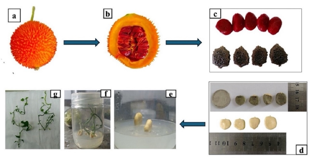
The hypocotyl region was selected as the explant source because preliminary observations indicated that this tissue showed an early and consistent callogenesis response compared with other seedling parts. This response may be associated with the juvenile nature of hypocotyl tissue, its relatively high meristematic activity, and its sensitivity to exogenous auxin and cytokinin combinations, which may facilitate dedifferentiation and callus initiation under in vitro culture conditions. The progression from mature fruit, seed without arillus, and seed culture to four week old in vitro seedlings is shown in
Fig. 1.
Callus induction was carried out using hypocotyl explants from four-week-old Dumbaya seedlings cultured on MS medium supplemented with different combinations of 2,4-D and 6-BA for eight weeks. Visible callus formation was observed in all tested treatments. All hypocotyl explants cultured in A1, A2, and A3 successfully formed callus, resulting in a 100% callus induction frequency across all treatments. Although callus induction frequency was uniform, callus color and browning degree varied among treatments. The induced callus was generally white to yellowish white, with some tissues gradually turning brown toward the end of the culture period. The predominant callus texture was friable, indicating a loose and crumbly structure. A summary of callus induction frequency, color, and texture for each treatment is presented in
Table 2.
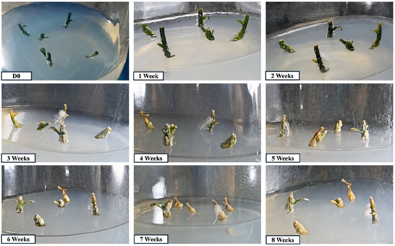
Fig. 2 shows the development of callus growth under treatment A1 from the day of inoculation (D0) to 8 Weeks. At the D0, most of the explants still appeared fresh and green; however, one explant had already shown callus formation. This callus was not induced by treatment A1 but had formed earlier during the seed germination stage until the development of Dumbaya seedlings. Therefore, the presence of callus at D0 can be considered pre-existing or initial callus originating from the pre-treatment stage. At 1 Week, the explants began to adapt to the culture medium. Several explants still maintained their green coloration, while the basal parts or wounded areas began to show slight swelling. At 2 weeks, a morphogenetic response became visible, indicated by the emergence of roots in several explants, particularly from the basal parts in contact with the medium. This suggests that treatment A1 began to stimulate an early growth response, especially root formation. At 3-4 weeks, root growth became more evident, accompanied by the appearance of newly formed callus tissue on several parts of the explants, particularly in the basal or wounded areas. At 5-6 Weeks, callus growth became more pronounced, with the callus masses appearing larger and showing a friable texture. The observed callus color under A1 treatment was predominantly white and brown. The growth response among explants was not uniform, indicating variation in the tissue regeneration capacity of each explant. At 7-8 Weeks, the callus masses became more visible, and brown coloration was increasingly observed in some tissues. This brown coloration may indicate tissue browning or reduced viability in some explants.
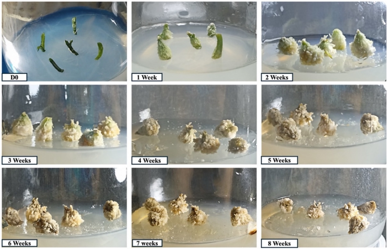
Under the A2 treatment,
Fig. 3 shows progressive morphological changes in Dumbaya hypocotyl explants during 8 weeks culture period. At the initial stage of culture (D0), the explants remained green and intact on the culture medium. By 1 week tissue swelling was observed, indicating an early response to callus induction. At 2-3 weeks, callus formation became clearly visible on the explant surface, particularly around the basal and wounded regions. From 4 to 6 weeks, the callus increased in size and volume, showing a friable, granular, and irregular morphology. The observed callus color under A2 treatment was predominantly white and brown. At 7-8 weeks, the callus masses became more developed, although brown coloration was observed in several regions, which may be associated with tissue browning, aging, or oxidative responses. Overall, the A2 treatment effectively induced progressive callus proliferation in Dumbaya hypocotyl explants, producing friable callus with white and brown coloration.
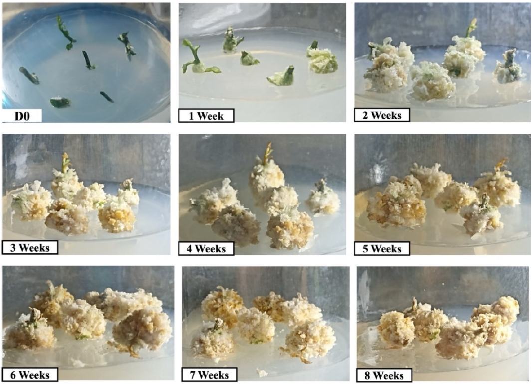
Treatment A3 produced a yellowish white friable callus with less visible browning (
Fig. 4). During 8 weeks culture period, explants showed gradual morphological changes. At D0 and 1 Week, green hypocotyl tissues were still visible, and early callus formation appeared at the cut surfaces. By 2-3 Weeks, newly formed callus masses appeared mainly white to yellowish white, with small patches of green tissue remaining. From 4 to 6 weeks, the callus increased in volume and began to show scattered brown areas, suggesting tissue aging or oxidation. At 7 and 8 weeks, the callus remained predominantly white to yellowish white, although browning became more visible in older outer regions.
The estimated callus fresh weight was measured at eight weeks after culture initiation using a digital balance. Because fresh weight was calculated indirectly based on the difference between the final and initial weights of culture bottles containing medium and callus, the values are presented as estimated fresh weight. The estimated fresh weight of callus under different growth regulator treatments is presented in
Table 3.
The highest mean estimated fresh weight was observed in A3 (0.80 ± 0.46 g), followed by A2 (0.53 ± 0.06 g) and A1 (0.27 ± 0.21 g). However, the Kruskal-Wallis test showed that the differences among treatments were not statistically significant, H(2)=3.586, p=0.166. Therefore, A3 should be interpreted as showing a descriptive trend toward higher estimated fresh weight rather than a statistically superior response.
A supplementary one-way ANOVA showed a similar non-significant pattern, F=2.494,
p=0.163 (
Table 4). Thus, under the culture conditions used in this study, the tested combinations of 2,4-D and 6-BA did not significantly affect the estimated fresh weight of Dumbaya callus.
Discussion
In vitro Seed Based Regeneration and Aseptic Seedling Production
The results of this study showed that Dumbaya seeds could be established under in vitro conditions on hormone- free MS medium. The successful development of aseptic seedlings is an important preliminary step because these seedlings provide the hypocotyl explants used for callus induction. A stepwise sterilization procedure using 5% Clorox and 70% ethanol, followed by rinsing with sterile distilled water and inoculation under aseptic conditions in a laminar airflow cabinet, was effective in producing sterile seeds capable of germinating on MS0 medium at approximately 25-26℃.
This finding is relevant because contamination remains one of the major constraints in seed-based in vitro culture systems, particularly in medicinal plants. For example, contamination has been reported to reduce the efficiency of in vitro culture establishment in
Nigella damascena (
Klimek-Chodacka et al. 2020). The successful germination of Dumbaya on basal MS medium indicates that this species is responsive to basic in vitro culture conditions and that seed-derived aseptic seedlings can serve as a reliable explant source for subsequent callus induction. Similar approaches have also been emphasized in the tissue culture systems of
Bupleurum chinense and
Tussilago farfara, where healthy aseptic seedlings are important for further morphogenic responses (
Bojko et al. 2024;
Li et al. 2023).
Hypocotyl segments from four-week-old in vitro seedlings were responsive to callus induction. Visible callus formation was observed in all tested treatments, and all cultured hypocotyl explants successfully formed callus. Thus, the callus induction frequency reached 100% in A1, A2, and A3. This result indicates that all tested combinations of 2,4-D and 6-BA were capable of inducing callus from Dumbaya hypocotyl explants under the present culture conditions.
The responsiveness of hypocotyl explants may be related to the juvenile nature of this tissue. Young hypocotyl tissues generally contain actively dividing cells and may be more easily reprogrammed into proliferative callus cells than mature tissues. Similar responses have been reported in other plant species, where hypocotyl explants were effective for callus induction and regeneration-related processes, including
Nigella damascena, Physalis angulata, Bupleurum chinense, and
Baccaurea angulata (
Klimek-Chodacka et al. 2020;
Li et al. 2023;
Mastuti et al. 2020;
Teresia et al. 2024). The higher responsiveness of young tissues is also consistent with the concept that cells with higher developmental plasticity are more readily redirected toward callus formation under appropriate hormonal conditions (
Kruglova et al. 2023). However, because this study did not compare hypocotyls with other explant types, the relative superiority of hypocotyl explants in Dumbaya should be confirmed in future comparative studies.
Callus induction from Dumbaya hypocotyl explants was carried out on MS medium supplemented with three combinations of 2,4-D and 6-BA: A1, 2 mg L
-1 2,4-D + 0.5 mg L
-1 6-BA; A2, 1.5 mg L
-1 2,4-D + 0.75 mg L
-1 6-BA; and A3, 1 mg L
-1 2,4-D + 1.5 mg L
-1 6-BA. Callus formation in all treatments supports the role of auxin and cytokinin interaction in promoting dedifferentiation and cell proliferation. 2,4-D is widely used to stimulate dedifferentiation and unorganized cell division, whereas 6-BA supports cell division and tissue growth. This response is consistent with classical and recent explanations of callus induction mechanisms, in which the balance between auxin and cytokinin regulates the transition of differentiated tissues into proliferative callus masses (
George et al. 1984;
Hendaryono et al. 1994;
Ikeuchi et al. 2013;
Pierik 1997;
Yin et al. 2024).
Although all treatments induced callus, differences were observed in callus color and visible browning intensity. Treatments A1 and A2 produced white to brown friable callus, whereas A3 produced yellowish-white friable callus with less visible browning. White or yellowish callus is generally associated with actively dividing tissue, whereas browning is commonly related to the accumulation and oxidation of phenolic compounds and tissue aging (
Jones et al. 2013;
Muna et al. 2022;
Sorentina et al. 2013). Therefore, the morphology observed in A3 suggests a more favourable descriptive response. However, this observation was qualitative and was not supported by quantitative color measurement; therefore, it should not be interpreted as definitive evidence of treatment superiority.
A comparable response has been reported in other in vitro systems, where suitable auxin and cytokinin combinations promoted vigorous callus growth, while less suitable conditions resulted in limited growth, browning, or early organogenic responses (
Palanivel et al. 2002;
Ren et al. 2017). In the present study, the lower visible browning in A3 may indicate that the balance between 2,4-D and 6-BA was more suitable for maintaining callus tissue in an actively proliferating state. Nevertheless, further validation using quantitative measurements of browning intensity, callus viability, and regeneration potential is required.
All tested treatments produced predominantly friable callus. Callus texture is commonly classified as compact, intermediate, or friable, and these differences are influenced by explant type, medium composition, plant growth regulator concentration, and culture environment (
Pierik 1997;
Turhan et al. 2004). The interaction between wounded explant tissue and the culture medium can stimulate swelling, cell proliferation, and callus formation in response to both injury and exogenous plant growth regulators (
Purba et al. 2017;
Rasud et al. 2020).
The predominance of friable callus in Dumbaya is relevant because friable callus is generally easier to separate into smaller cell aggregates and may be useful for subsequent culture manipulation. Similar findings have been reported by
Kurniati (2013), who observed friable and compact callus formation from hypocotyl explants of white chili (
Capsicum frutescens), and by
Mastuti et al. (2020), who reported friable callus formation from ciplukan (
Physalis angulata) hypocotyls.
During the eight week culture period, callus morphology changed gradually. Early callus development was observed at the cut surfaces, followed by increased callus mass and a transition toward friable yellowish white tissue. Under A3, the callus showed less visible browning and a more consistent friable appearance. However, because visual morphology was assessed descriptively, this response should be interpreted as a qualitative trend rather than definitive evidence that A3 was statistically superior to the other treatments.
Callus Fresh Weight
The estimated callus fresh weight was used as a quantitative indicator of biomass accumulation because fresh weight is commonly used to describe callus growth performance in in vitro culture systems (
Palanivel et al. 2002;
Shah et al. 2020;
Waryastuti et al. 2017). In this study, fresh weight was estimated indirectly based on the difference between the final and initial weights of culture bottles containing medium and callus. Therefore, the data should be interpreted as estimated biomass accumulation rather than direct fresh weight measurement.
Treatment A3 produced the highest mean estimated fresh weight (0.80 ± 0.46 g), followed by A2 (0.53 ± 0.06 g) and A1 (0.27 ± 0.21 g). Descriptively, this pattern suggests that the combination of lower 2,4-D and higher 6-BA in A3 tended to support greater callus biomass accumulation. This is consistent with previous reports showing that plant growth regulator composition can influence callus proliferation and biomass formation across different plant species (
Noli 2015;
Waryastuti et al. 2017).
However, the descriptive increase observed in A3 was not statistically significant. The Kruskal-Wallis test showed no significant difference in estimated callus fresh weight among treatments, H(2)=3.586, p=0.166. A supplementary one-way ANOVA also showed no significant effect of treatment on estimated fresh weight, F=2.494, p=0.163. Therefore, A3 should not be interpreted as a statistically superior treatment. Instead, A3 may be considered a promising candidate formulation because it showed a descriptive trend toward higher estimated fresh weight, yellowish-white friable callus morphology, and lower visible browning.
The relatively large standard deviation in A3 (0.80 ± 0.46 g) indicates biological variation among explants in response to the treatment. Such variation may reduce statistical power and make it difficult to detect significant differences among treatments, especially when the number of replications is limited. Similar issues have been reported in callus culture studies where visual or descriptive trends in biomass accumulation were not always supported by statistically significant differences due to biological variability or limited replication (
Kumar et al. 2008;
Pajević et al. 2004;
Palanivel et al. 2002;
Silva et al. 2005).
The temporal pattern of callus development from 1 - 8 weeks showed that Dumbaya callus underwent gradual changes in color and texture. Initially, the callus appeared white to green and showed a mixed compact-friable texture. As the culture period progressed, the callus became increasingly friable and yellowish. This pattern may indicate continued cell proliferation and biomass accumulation during the culture period.
Among the three treatments, A3 (1 mg L-1 2,4-D + 1.5 mg L-1 6-BA) showed a promising descriptive response based on several observed indicators, including a relatively higher mean estimated fresh weight, friable texture, yellowish white coloration, and lower visible browning. Nevertheless, because the differences in estimated fresh weight were not statistically significant, A3 should not be interpreted as a statistically superior treatment. Instead, A3 can be considered a preliminary candidate formulation for inducing friable callus from Dumbaya hypocotyl explants. Further optimization involving additional PGR concentrations, larger sample sizes, direct biomass measurement, and longer observation periods is needed to confirm the most reliable formulation for callus growth and stability.
The Future Perspectives and Study Limitations
Although the present study focused only on seed-based in vitro establishment, hypocotyl-derived callus induction, callus morphology, and estimated fresh weight, the results provide a preliminary basis for further research on Dumbaya tissue culture. The production of friable callus, particularly under treatment A3, may be useful for future studies aimed at developing regeneration protocols through organogenesis or somatic embryogenesis. However, these processes were not directly evaluated in the present study and should therefore be regarded as future research directions rather than conclusions from the current data.
Future work may also examine whether Dumbaya callus cultures are suitable for secondary metabolite-related studies. This is relevant because callus culture systems have been used in other medicinal and aromatic plants as controlled platforms for studying or producing bioactive compounds (
Boubertakh et al. 2013;
Demeter et al. 2010;
Gerdakaneh et al. 2011). Nevertheless, the present study did not analyze phytochemical content, metabolite profiles, or biosynthetic capacity. Therefore, any potential application of Dumbaya callus for secondary metabolite production remains hypothetical and requires direct experimental validation.
In addition, genetic and ecological variation among Dumbaya accessions should be considered in future studies. Previous reports have shown genetic variation among
M. cochinchinensis materials collected from different geographical locations (
Khairi et al. 2023;
Wimalasiri et al. 2016). Such variation may influence in vitro responses, including callus induction and biomass accumulation. However, because the present study did not assess genetic diversity or compare multiple accessions, this factor cannot be used to explain the observed results conclusively. Future studies involving different Dumbaya accessions are required to determine whether genetic background affects callus induction efficiency and callus growth characteristics.
The present study also has several methodological limitations. First, only three combinations of 2,4-D and 6-BA were tested, and no PGRs free control medium was included for callus induction. Second, the replication level was limited, which may affect statistical reliability. Third, callus fresh weight was estimated indirectly using bottle weight differences, which may introduce measurement error. Therefore, future studies should include direct measurement of callus fresh and dry weight, quantitative assessment of callus induction frequency, time to callus initiation, browning percentage, and regeneration capacity.
This study demonstrated that hypocotyl explants derived from four-week-old in vitro seedlings of M. cochinchinensis were responsive to callus induction on MS medium supplemented with 2,4-D and 6-BA. All tested treatments induced friable callus, with callus formation observed in all cultured explants. Treatment A3, consisting of 1 mg L-1 2,4-D and 1.5 mg L-1 6-BA, showed a descriptive trend toward higher estimated fresh weight and more favorable yellowish white friable callus morphology with lower visible browning. However, the differences in estimated fresh weight among treatments were not statistically significant. Therefore, A3 should be considered a preliminary candidate formulation rather than a statistically confirmed optimal treatment. Further studies using a PGRs free control, additional growth regulator combinations, larger sample sizes, direct biomass measurement, and quantitative assessment of callus initiation and regeneration capacity are needed to validate and optimize the callus induction protocol for M. cochinchinensis.
Notes
-
Acknowledgments
The authors would like to express their sincere gratitude to the community of Ilomata Village, Atinggola District, North Gorontalo Regency, Indonesia, for their invaluable support and for preserving the traditional use of herbal plants for medicinal purposes. This work was supported by the Directorate General of Higher Education, Research, and Technology through the DRTPM-BIMA Program under contract number 935/UN47.D1.1/PT.01.03/2024, within the Regular Fundamental Research Scheme.
Fig. 1.Stages of Dumbaya seed germination. (a) Fresh, fully ripe Dumbaya fruit; (b) longitudinal section of the fruit after being split open; (c) seed morphology from seeds still covered by the arillus (Pagalla et al. 2023); (d) the seeds without hard seed coat; (e) seeds cultured on MS medium without PGRs; (f, g) germination results at 4 weeks after planting.
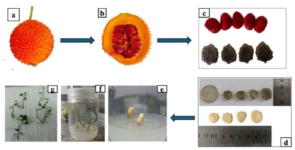
Fig. 2.Development of callus growth in Dumbaya hypocotyl explants under A1 treatment from the day of inoculation (D0) to 8 Weeks. The callus produced under this treatment was characterized by white and brown coloration with a friable texture.
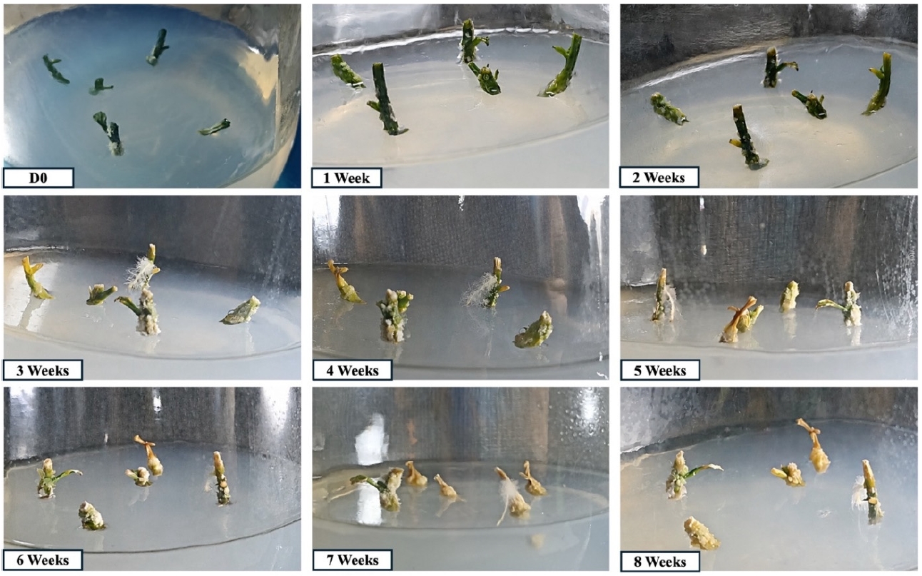
Fig. 3.Morphological changes during callus induction from Dumbaya hypocotyl explants under A2 treatment from the day of inoculation (D0) to 8 weeks. The callus produced under this treatment was characterized by white and brown coloration with a friable texture.
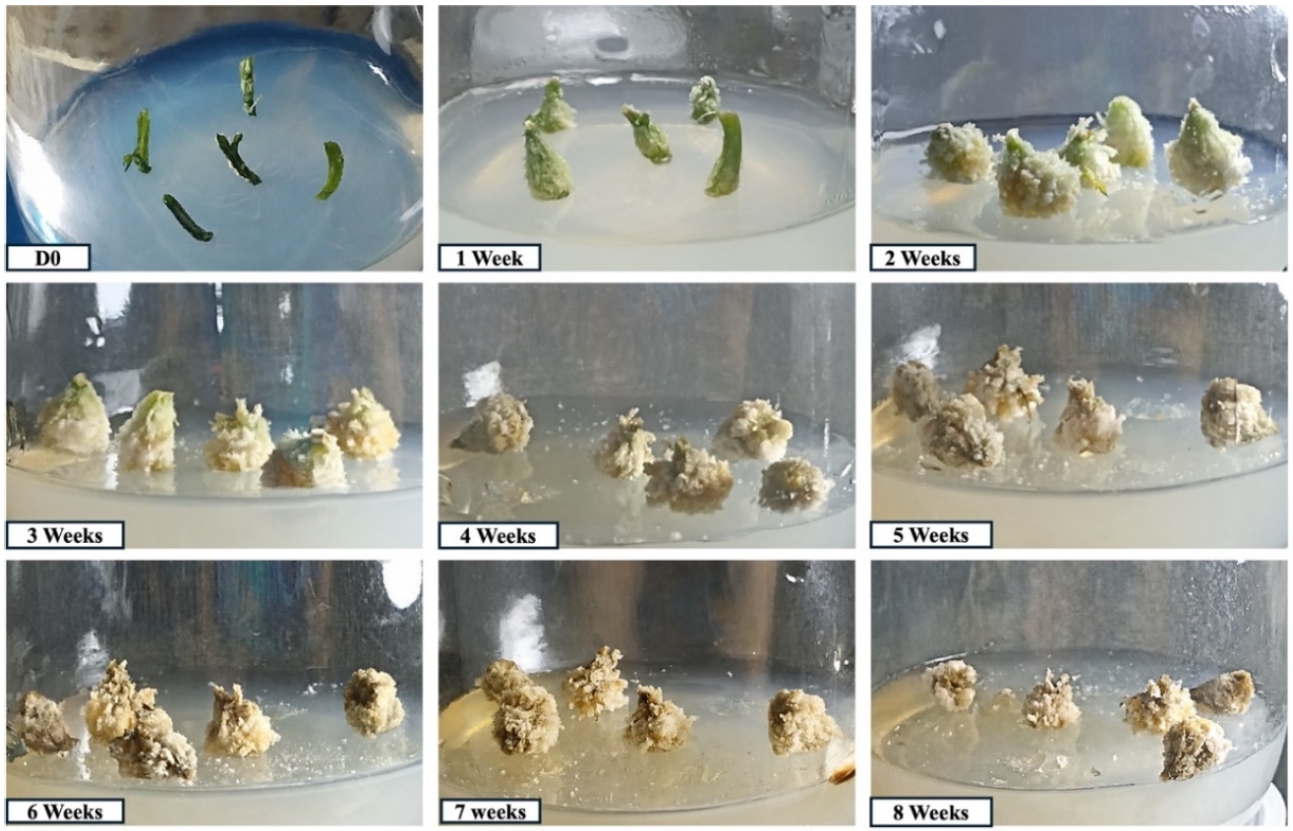
Fig. 4.Morphological changes during callus induction from Dumbaya hypocotyl explants, from the time of inoculation (D0) to 8 weeks.
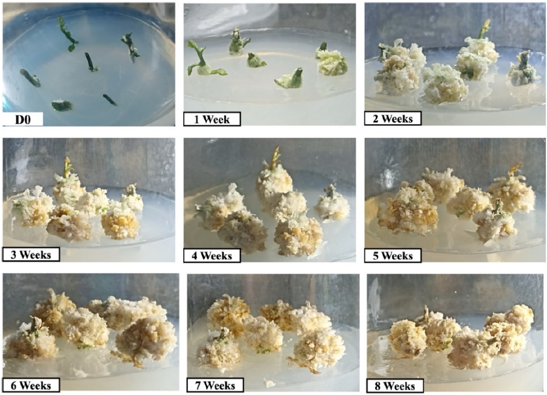
Table 1.
Table 1.
|
Label |
Treatment |
|
A1 |
MS + 2.0 mg L-1 2,4-D + 0.5 mg L-1 6-BA |
|
A2 |
MS + 1.5 mg L-1 2,4-D + 0.75 mg L-1 6-BA |
|
A3 |
MS + 1.0 mg L-1 2,4-D + 1.5 mg L-1 6-BA |
Table 2.Callus induction frequency, color, and texture of Dumbaya callus after 8 weeks of culture.
Table 2.
|
Treatment |
Explant |
Callus induction frequency (%) |
Color |
Texture |
|
A1 |
Hypocotyl |
100 |
White, brown |
Friable |
|
A2 |
Hypocotyl |
100 |
White, brown |
Friable |
|
A3 |
Hypocotyl |
100 |
Yellowish white |
Friable |
Table 3.The effect of different treatments on fresh weight callus.
Table 3.
|
Treatment |
n |
Fresh weight (g), mean ± SD |
Median |
Min-Max |
Mean rank |
|
A1 |
3 |
0.27 ± 0.21 |
0.20 |
0.10-0.50 |
2.67 |
|
A2 |
3 |
0.53 ± 0.06 |
0.50 |
0.50-0.60 |
5.67 |
|
A3 |
3 |
0.80 ± 0.46 |
0.70 |
0.40-1.30 |
6.67 |
|
Kruskal-Wallis test |
|
|
|
|
H(2)=3.586; p=0.166 |
Table 4.The one-way ANOVA of callus fresh weight among treatments.
Table 4.
|
Source of variation |
Sum of squares |
df |
Mean square |
F |
Sig. |
|
Between treatments |
0.427 |
2 |
0.213 |
2.494 |
0.163 |
|
Within treatments |
0.513 |
6 |
0.086 |
|
|
|
Total |
0.940 |
8 |
|
|
|
References
- Andaryani S, Samanhudi YA, Yunus A. 2019. Effect of BAP and 2, 4-D on callus induction of Jatropha curcas in vitro. Cell Biol Dev. 3(2): 56-65.
- Anjani DD. 2023. The effect of BAP and NAA combination on callus induction of aglaonema siam aurora leaf explants in vitro. Indonesian Journal of Bioscience (IJOBI). 1(2): 85-92.
- Bidabadi SS, Jain SM. 2020. Cellular, molecular, and physiological aspects of in vitro plant regeneration. Plants. 9(6): 702.
- Bojko M, Kędra M, Adamska A, Jakubowska Z, Tuleja M, Myśliwa-Kurdziel B. 2024. Induction and characteristics of callus cultures of the medicinal plant Tussilago farfara L. Plants. 13(21): 3080.
- Boubertakh B, Liu X-G, Cheng X-L, Li P. 2013. A spotlight on chemical constituents and pharmacological activities of Nigella glandulifera Freyn et Sint seeds. J. Chem. 2013(1): 820183.
- Demeter Z, Surányi G, Molnár VA, Sramkó G, Beyer D, Kónya Z, et al. 2010. Somatic embryogenesis and regeneration from shoot primordia of Crocus heuffelianus. Plant Cell Tissue Organ Cult. 100(3): 349-353.
- Febriani PF, Susanti R, Nazreena OA, Ab RZ. 2025. Induction and regeneration of ginger callus (Zingiber officinale) in Malaysia by Administration of 2, 4 D and NAA. Jurnal Agronomi Tanaman Tropika (JUATIKA). 7(1): 98-107.
- George E, Sherrington P. 1984. Plant propagation by tissue culture eastern press. England. 555(55): 55.
- Gerdakaneh M, Mozafari A-A, Sioseh-mardah A, Sarabi B. 2011. Effects of different amino acids on somatic embryogenesis of strawberry (Fragaria× ananassa Duch.). Acta Physiol. Plant. 33(5): 1847-1852.
- Hendaryono IDPS, Wijayani IA. 1994. Teknik kultur jaringan, pengenalan dan petunjuk perbanyakan tanaman secara vegetatif-modern. Kanisius.
- Hussain MS, Fareed S, Ansari S, Rahman MA, Ahmad IZ, Saeed M. 2012. Current approaches toward production of secondary plant metabolites. J. Pharm. Bioallied Sci. 4(1): 10-20.
- Ikeuchi M, Sugimoto K, Iwase A. 2013. Plant callus: Mechanisms of induction and repression. Plant Cell. 25(9): 3159-3173.
- Iswanto, A’ida N, Wahyuningsih, Restu M, Larekeng SH, Gusmiaty. 2025. Induction callus of species Palapi (Heritiera javanica) in several combinations of concentrations of 2, 4 Dichlorophenoaccetate and BAP. Presented at IOP Conference Series: Earth and Environmental Science.
- Jones AMP, Saxena PK. 2013. Inhibition of phenylpropanoid biosynthesis in Artemisia annua L.: A novel approach to reduce oxidative browning in plant tissue culture. PloS One. 8(10): e76802.
- Khairi M, Othman M. 2023. Morphological and genetic analysis of momordica cochinchinensis (Lour.) Spreng. (Gac) from different accessions in Malaysia. J. Trop. Life Sci. 13(2): 319.
- Klimek-Chodacka M, Kadluczka D, Lukasiewicz A, Malec-Pala A, Baranski R, Grzebelus E. 2020. Effective callus induction and plant regeneration in callus and protoplast cultures of Nigella damascena L. Plant Cell Tissue Organ Cult. 143(3): 693-707.
- Kruglova N, Zinatullina A, Yegorova N. 2023. Histological approach to the study of morphogenesis in callus cultures in vitro: a review. Int. J. Plant Biol. 14(2): 533-545.
- Kumar N, Pamidimarri SD, Kaur M, Boricha G, Reddy MP. 2008. Effects of NaCl on growth, ion accumulation, protein, proline contents and antioxidant enzymes activity in callus cultures of Jatropha curcas. Biologia. 63(3): 378-382.
- Kurniati E. 2013. Induksi kalus dan penghasilan capsaicin pada variasi kadar nutrien MS dan kombinasi zat pengatur tumbuh. UAJY.
- Li Y, Zhao J, Chen H, Yu X, Li H, Zhang Y, et al. 2023. Plant regeneration via callus-mediated organogenesis in commercial variety of Chuanbeichai No. 1 in Bupleurum chinense DC. Plant Biotechnol. Rep. 17(2): 159-169.
- Mastuti R, Widoretno W, Harijati N. 2020. Kultur kalus tanaman obat ciplukan (Physalis angulata L.). Biotropika. 8(1): 26-35.
- Muna A, Suharyanto S, Sasongko AB. 2022. Induksi kalus Piper retrofractum Vahl. Dengan variasi eksplan dan zat pengatur tumbuh. Quagga: Jurnal Pendidikan dan Biologi. 14(1): 16-23.
- Muthi’ah A, Sakya A, Setyawati A, Samanhudi, Rahayu M. 2023. Callus induction of Calotropis gigantea using BAP and 2, 4-D in vitro. Presented at IOP Conference Series: Earth and Environmental Science.
- Noli ZA. 2015. Induksi kalus Artemisia vulgaris L. dengan Pemberian Beberapa Konsentrasi 2, 4-Dichlorophenoxyacetic Acid (2, 4-D). Jurnal Biologi UNAND. 4(4): 216-222.
- Pagalla DB, Ahmad J, Adudu MF, Nidaulhasanah A, Adju FHY, Damayanti EM. 2023. In vitro germination of dumbaya seeds (Momordica cochinchinensis (Lour.) spreng: A unique medicinal plant of gorontalo. J. Biol. Tropis. 23(2): 203-208.
- Pajević S, Vasić D, Sekulić P. 2004. Biochemical characteristics and nutrient content of the callus of sunflower inbred lines/características bioquímicas y el contenido de nutrientes en los callos de las líneas consanguíneas (inbred) de girasol/caractéristiques biochimiques et contenu de nutriment dans les callus des lignées de tournesol. Helia. 27(41): 143-150.
- Palanivel S, Parvathi S, Jayabalan N. 2002. Callus induction and plantlet regeneration from mature cotyledonary segments of groundnut (Arachis hypogaea L.). J. Plant Biol. 45(1): 22-27.
- Pierik RLM. 1997. In vitro culture of higher plants. Springer Science & Business Media.
- Purba RV, Yuswanti H, Astawa ING. 2017. Induksi kalus eksplan daun tanaman anggur (Vitis vinivera L.) dengan aplikasi 2, 4-D secara in vitro. E-Jurnal Agroekoteknologi Tropika. 6(2): 218-228.
- Rasud Y, Bustaman B. 2020. Induksi kalus secara in vitro dari daun cengkeh (Syizigium aromaticum L.) dalam media dengan berbagai konsentrasi auksin. Indones. J. Agric. Sci. 25(1): 67-72.
- Ren J-W, Lei Y, Li X-L. 2017. Tissue culture of callus and establishment of regeneration system of Tussilago farfara petiole. Zhongguo Zhong yao za zhi= Zhongguo Zhongyao Zazhi= China Journal of Chinese Materia Medica. 42(20): 3895-3900.
- Shah M, George IA. 2020. Pigment elicitation and sun protection factor of callus induced from Cassia tora seedling explants. Plant Cell Tissue Organ Cult. 143(1): 201-210.
- Silva ALCd, Caruso CS, Moreira RdA, Horta ACG. 2005. Growth characteristics and dynamics of protein synthesis in callus cultures from Glycine wightii (Wight & Arn.) Verdc. Ciênc. Agrotecnologia. 29(6): 1161-1166.
- Sorentina MS, Haliani M, Suwastika IN. 2013. Induksi kalus bawang merah (Allium ascalonicum L.) lokal palu pada medium MS dengan penambahan 2, 4-D (2, 4-asam dikloropenoksi asetat) dan air kelapa. J. Nat. Sci. 2(2): 55-63.
- Teresia N, Zakiah Z, Turnip M. 2024. Induksi kalus dari hipokotil belimbing merah (Baccaurea angulata) dengan penambahan 2, 4-D (Dichlorophenoxy acetic acid) dan BAP (6-Benzyl Amino Purin). J. Biol. Tropis. 24(1): 194-203.
- Turhan H, Baser I. 2004. Callus induction from mature embryo of winter wheat (Triticum aestivum L.). Asian J. Plant Sci. 3(1): 17-19.
- Ummah M, Mashluhah K, Resmisari R. 2020. Conservation of juwet (Syzygium cumini) plant using in vitro culture techniques. EL-HAYAH. 7(3): 106-112.
- Waryastuti DE, Setyobudi L, Wardiyati T. 2017. Pengaruh tingkat konsentrasi 2, 4-D dan BAP pada media MS terhadap induksi kalus embriogenik temulawak (Curcuma xanthorrhiza Roxb.). Brawijaya University.
- Wimalasiri D, Piva T, Urban S, Huynh T. 2016. Morphological and genetic diversity of Momordica cochinchinenesis (Cucurbitaceae) in Vietnam and Thailand. Genet. Resour. Crop Evol. 63(1): 19-33.
- Yin R, Chen R, Xia K, Xu X. 2024. A single-cell transcriptome atlas reveals the trajectory of early cell fate transition during callus induction in Arabidopsis. Plant Commun. 5(8): 100941.